Cerebral Palsy (CP) is known as a group of disorders that affect the development of posture and movement which cause limitations in activity. CP has been attributed to non-progressive disturbances that occurred during the development of the fetal or infant brain. Such disturbances can include but are not limited to; preterm birth, multiple gestation, intrauterine growth restriction, prenatal stroke, maternal iodine deficiency, and intrauterine infections. Cerebral Palsy is limited to lesions in the brain only. These lesions can occur anywhere between fetal development and 3 years of age. Approximately 30-50% of patients with CP also have mental retardation and approximately 15-60% of children with CP have epilepsy. There is currently no cure for CP, but with the right therapy patients may become better integrated and more independent.
MRI is currently the best known imaging modality to diagnose cerebral palsy because it shows cortical and white matter structures and abnormalities more clearly than any other imaging modality. Most often MRI is used to determine the cause of spasiticity(muscles tightness) and can detect brain elongation and bleeding which are both early signs of CP. MRI is also used when imaging patients who have already been diagnosed with cerebral palsy because it can help determine the extent of damage CP has already done on the patients body.
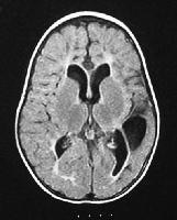
MRI of a 16 month old male who was born at term but had an anoxic event at delivery. The image shows asymetric cerebral palsy more prominent on the right side. Decreased white matter and enlarged ventricles seen in this image are indicative of Cerebral Palsy.