Wednesday, April 13, 2011

Trigeminal Neuralgia

Trigeminal neuralgia(TN) is a disorder of the 5th cranial nerve that causes stabbing or electric shock like pain in parts of the face.  This pain comes directly from the trigeminal nerve in the face and can affect all of the face and part of the eye.  The trigeminal nerve is responsible for carrying pain, feelings and other sensations from the brain to the parts of the face.  This condition does not specifically target any certain age or race and maybe be considered a normal part of the aging process.  Trigeminal neuralgia most often occurs due to increased pressure on the trigeminal nerve from a blood vessel or tumor, but can also be caused by multiple sclerosis.  The most common symptom is very painful or sharp electric feeling pain that lasts a few minutes but may become constant.  Pain maybe triggered by light touch or stimulus and usually only occurs on one side of the face around the eye, cheek and lower part of the face.  Often times the painful experience arises after trauma to the face or neck, such as whiplash.
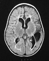
To fully diagnose trigeminal neuralgia a variety of exams are performed such as a neurological exam.  MRI is the typical imaging study used to diagnose trigeminal neuralgia.  MRI will most often reveal a slow-growing brain tumor compressing the nerve.  Treatment options do exist and have proven to be successful in reducing the amount of attacks and the severity of pain.  Treatment options include anti-convulsive drugs or surgery.


                    The three areas the trigeminal nerve branches out to in which the patient will feel pain.


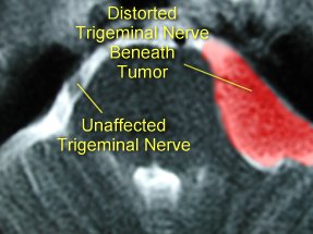
MRI axial image of the trigeminal nerve being compressed beneath a tumor causing TN compared to a normal trigeminal nerve.

Saturday, April 9, 2011

Cherubism

Cherubism is a disease characterized by abnormal bone tissue in the lower part of the face.  Both the mandible and maxilla become enlarged and the bone is replaced with cyst-like growths which are painless.  These growths give the cheeks a swollen, round appearance and often times interfere with tooth development.  Cherubism is a hereditary disease affecting the genes.  The first signs of cherubism are usually seen in early childhood and continue to grow until puberty.  The abnormal growths are gradually replaced with normal bone in early adulthood which is why most affected adults have a normal facial appearance.  Cherubism rarely affects any other parts of the body unless it occurs as part of another genetic disorder, such as Ramon syndrome.
In imaging studies cherubism is similar to fibrous dysplasia especially when the excessive growth is confined to the mandible.   Because of the similarities it is suggested that cherubism may be a form of fibrous dysplasia.  Imaging procedures used to diagnose cherubism include CT scans and MR imaging. 
Cherubism seen in a young girl with excessive growth of the mandible and maxilla.

                                       
3D Coronal oblique CT scan of a 16 year old girl with massive mandibular enlargement and difficulty swallowing.
Coronal CT scan of 16 year old girl with massive mandibular enlargement shows variable appearance of matrix, ranging from radiolucent to densely sclerotic.

Friday, March 25, 2011

Colobomas

A coloboma is a congenital malformation in which part of the eye does not form due to failure of fusion of embryonic structure called the intraocular fissure.  This malformation can occur unilaterally or bilaterally and causes the eye to protrude anteriorly or posteriorly.  There are many places a coloboma can occur; the iris, the lens, the choroid, the eyelid, the optic disc, or the retina.  Colobomas are present at birth and usually affect 1 out of every 10,000 births.  A coloboma can occur due to surgery, trauma or may be hereditary.   If a child is diagnosed with a coloboma it is suggested that the parent have an eye exam because this condition can be present without any vision problems. The effect this condition has on vision depends on where in the eye the coloboma is located and how big the gap is. One example of a vision defect is a dislike to bright lights, which usually occurs in those with a coloboma in the iris.   The key finding in children with this condition is a key-hole shaped pupil and may be diagnosed with the help of an ophthalmologist and MRI exam.  A coloboma is typically identified on an MRI T2 weighted image as a hypodense protrusion of the orbital globe. Children affected by colobomas sometimes have an accompanying rare condition called CHARGE syndrome, which stand for; coloboma, heart defects, atresia of the choanae (problems with the nose passage), retarded growth and development, genital hypoplasia (undescended testicles), ear abnormalities.   Currently there is no treatment for colobomas, except that the child will have help from a specialist to determine the effects and problems of the coloboma.  Most children will be required to have eye exams every six months until the age of seven and then yearly after the age of seven, and will be prescribed sunglasses to help with light sensitivity.
                              Pediatric MRI T2 weighted image of a coloboma on the right side.

                                           Key-hole shaped pupil in a child with a coloboma.

Sunday, March 20, 2011

Acromegaly

Acromegaly is a tumor of the pituitary gland that causes increased thickening of the bones in the hands, feet, cheeks and jaw.  Acromegaly occurs in about 6 of every 100,000 adults and is usually caused by the production of excessive growth hormone after the skeleton is finished growing.  This overproduction of growth hormone is a result of a benign tumor of the pituitary gland.  Some common symptoms include but are not limited to; impaired vision, body odor, carpal tunnel syndrome, decreased muscle strength, easy fatigue, headache, excessive height, enlarged hands, feet, jaw and tongue, limited joint movement, thickening of the skin, and excessive hair growth in females.  This disease is most often diagnosed in middle-aged adults, but can appear at any age.  Patients with acromegaly are at high risk for developing type II diabetes, hypertension, sleep apnea syndrome, cardiovascular disease, arthritis, and colon polyps which could develop into cancer if left untreated.
Acromegaly is usually diagnosed utilizing several different tests.  Two of the usual tests are the growth hormone blood test and oral glucose tolerance test to determine if the patient has excessive growth hormone in the body.  Likewise the physician also uses imaging modalities after growth hormone tests to determine location and size of the tumor.  The physician will most likely order an MRI of the pituitary gland.  The MRI technologist will utilize sagittal and coronal T1 weighted spin echo images before and after the use of gadolinium.   In most cases a macroadenoma (tumor bigger than 10mm) will be found.  In a gadolinium enhanced MRI acromegaly is usually seen as a hypodense area where the pituitary gland is located.
There are usually three approaches to treatment for acromegaly; surgery, medical therapy, and radiation therapy.  Surgery is usually the first option and usually has the best outcomes.  During surgery the surgeon simply removes the tumor, which relieves the pressure and allows the hormone levels to lower.  It is important to note that even after surgery there is a chance of reoccurrence so patients may need medications and may need to continue to be monitored.  Medical therapy uses three classes of drugs (SSAs, GHRAs, Dopamine antagonists) for treatment in patients who are unable to have surgery or to shrink a tumor prior to surgery.  Radiation therapy is used for patients who still have some tumor left after surgery or whose bodies are unresponsive to medications, although it is important to note the effects of radiation therapy may not be seen for years.

                     The picture above shows a common symptom, enlargement of the hands.


       MRI image of a macroadenoma in acromegaly.  The image on the right shows the tumor
is only about 9mm in size. The tumor is far enough away that there is no impairment of vision.

MRI image of pituitary macroadenoma in an acromegalic patient.  The picture on the right shows the tumor with a diameter of 30mm.  The optic chiasm is elevated and compressed causing the patient loss of peripheral vision.

Monday, March 14, 2011

Mesial Temporal Sclerosis

Mesial temporal sclerosis (MTS) is also known as hippocampal sclerosis.  This condition is closely related to temporal lobe epilepsy, which simply means that the start of the seizure can be identified within the temporal lobe of the brain.  Mesial temporal sclerosis is due to the loss of neurons and results in the scarring of the hippocampus due to certain brain injuries.  These brain injuries include but are not limited to infection, traumatic injury, tumors, and lack of oxygen to the brain.  Common symptoms include strange sensations, changes in behavior or emotions, muscle spasms, and convulsions.  MTS occurs in 65% of epilepsy patients in autopsy studies. MTS treatment usually includes anti-epileptic medications or temporal lobectomy.
Mesial temporal sclerosis is best viewed using MRI.  The best imaging planes to detect MTS are the coronal plane and the axial plane.  The coronal plane is able to detect volume reduction and T2 signal change while the axial plane is able to detect the posterior extent of hippocampal abnormalities.  Likewise gradient echo techniques are used rather than spin echo due to the smaller slice thickness.  Because gradient echo allows for smaller slice thickness it allows for better imaging of gray and white matter.  On a typical MRI study showing MTS you may find the following abnormalities; reduced hippocampal volume, increased T2 signal, loss of gray and white matter interface in the anterior temporal lobe, atrophy of the ipsilateral fornix and mammillary body.
Above is a video showing MRI images of MTS along with a short clip of a left mesial temporal sclerosis with calcified lesion removal.

Monday, February 21, 2011

Cerebral Palsy and MRI

Cerebral Palsy (CP) is known as a group of disorders that affect the development of  posture and movement which cause limitations in activity.  CP has been attributed to non-progressive disturbances that occurred during the development of the fetal or infant brain. Such disturbances can include but are not limited to; preterm birth, multiple gestation, intrauterine growth restriction, prenatal stroke, maternal iodine deficiency, and intrauterine infections. Cerebral Palsy is limited to lesions in the brain only. These lesions can occur anywhere between fetal development and 3 years of age. Approximately 30-50% of patients with CP also have mental retardation and approximately 15-60% of children with CP have epilepsy.  There is currently no cure for CP, but with the right therapy patients may become better integrated and more independent.

MRI is currently the best known imaging modality to diagnose cerebral palsy because it shows cortical and white matter structures and abnormalities more clearly than any other imaging modality. Most often MRI is used to determine the cause of spasiticity(muscles tightness) and can detect brain elongation and bleeding which are both early signs of CP.  MRI is also used when imaging patients who have already been diagnosed with cerebral palsy because it can help determine the extent of damage CP has already done on the patients body.


                                                MRI of a 16-month-old boy who was born at term bu...
MRI of a 16 month old male who was born at term but had an anoxic event at delivery.  The image shows asymetric cerebral palsy more prominent on the right side.  Decreased white matter and enlarged ventricles seen in this image are indicative of Cerebral Palsy.